The Sensitivity Test of Mycobacterium tuberculosis to Snail Seromucoid and Chitosan in vitro
Abstract
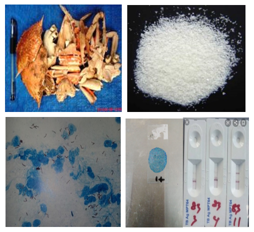
Tuberculosis (TB) is an infection caused by M. tuberculosis (MTb) and is transmitted through droplets of phlegm in the air from patients or those suspected of having TB. In general, treatment for TB is done with anti-tuberculosis drugs (ATDs), specifically streptomycin, isoniazid, rifampicin, and ethambutol (SIRE) that takes a long time due to the level of resistance of MTb bacteria. The resistance of MTb triggers ATDs based on natural bioactive compounds. Chitosan as a result of chitin deacetylation can function as an antimicrobial agent because it is polycationic, which is biodegradable, biocompatible, and non-toxic. Snail (Achatina fulica) seromucoid contains antibacterial bioactive compounds, namely glycans, peptides, glycopeptides, achasin protein, and chondroitin sulfate. This study aims at testing the sensitivity of MTb isolates against snail seromucoid and chitosan in vitro. This research applied the experimental research method. MTb isolates were obtained from sputum samples of patients suspected of TB at the Surakarta Regional Public Hospital (RSUD Surakarta). The results of screening for MTb were positive, based on the microscopic examination of MTb using the Ziehl Nelson (ZN) method, the MPT 64 rapid test, and the quick molecular test using the Genexpert method. The research was completed through several stages, including the preparation of a suspension of germs with a concentration of 1 mg/ml or Mc. Farland 0.5-1.0; preparation of the stock solution and working solution (WS); drug sensitivity test (DST) against snail seromucoid; chitosan and ATDs (SIRE) on Lowenstein Jensen (LJ) media; and incubation at 37°C for 3-4 weeks. The results were interpreted on day 28 or day 42. The results have revealed that MTb isolates are 100% resistant to snail seromucoid and 2% chitosan. This study concludes that MTb isolates from suspected TB are resilient to 100% snail seromucoid and 2% chitosan.
References
Bislimi, K. A. Behluli, J. Halili, I. Mazreku, F. Osmani, F. Halili (2013). Comparative Analysis of Some Biochemical Parameters in Emolymph of Garden Snail (Helix Pomatia L.) of The Kastriot and Ferizaj Regions, Kosovo. Dec 2013. Vol. 4, No. 6 pp 11 - 18 ISSN2305-8269; International Journal of Engineering and Applied Sciences © 2012 - 2013 EAAS & ARF www.eaas-journal.org
Burkovski, A. (2013). Cell Envelope of Corynebacteria: Structure and Influence on Pathogenicity. ISRN Mycrobiology, 013:11
Dolashka, P., Dolashki, A., Voelter, W., Van Beeumen, J., Stevanovic, S. (2015). Antimicrobial activity of peptides the hemolymph of Helix lucorum snails. International Journal of Current Microbiology and Applied Sciences ISSN: 2319-7706 Volume 4 Number 4(2015) pp. 1061-1071 http://www.ijcmas.com
Etim, L.B, Chuku, A. and Godwin, A.O. (2015). Antibacterial properties of snail mucus on bacteria isolated from patient with wound infection. British Microbiology Research Journal 11(2): 1-9, Article no. BNRJ.21731 ISSN: 2231-0886, DOI: 10.9734/BMRJ/2016/21731.
J. C. Fernandes, S. Humberto, A. S. Vanessa de Sousa, E. P. Manuela, and J. Francisco, “Anti-inflammatory activity of chito-oligosaccharides in vivo,” Marine Drugs Journal, vol. 8, pp. 1763-1768, 2010.
Harti, A.S., Nony P. Rahajeng P., 2019. Antimicrobial Bioactive Compounds of Snail Seromucoid as Biological Response Modifier Immunostimulator. Journal Microbiology Indonesia, Volume 13 edisi 2, Juni 2019. Print ISSN 1978-3477 Online ISSN 2087-8575 DOI 10.5454/mi.13.2.XX
Harti, A.S., Estuningsih, Kusumawati, H. N., Siswiyanti, Setyaningtyas, A. (2016). In vitro Synergistic Effect of Snail Slime and Chitosan against Staphylococcus aureus. International Journal of Pharma Medicine and Biological Science, ISSN: 2278-5221, Vol. 5 No 2 & No. 3, April 2016, pp. 137-141.
Harti, A.S., Sulisetyawati, S.D.,, Murharyati, A., Oktariani, M., & Wijayanti, I. B. (2016). The Effectiveness of Snail Slime and Chitosan in Wound Healing. International Journal of Pharma Medicine and Biological Science ISBN 2278-5221. Volume 5, Number 1, January 2016, page 76 – 80
Harti, A.S., Murharyati, A., Sulisetyawati, S.D., & Oktariani, M. (2018). The Effectiveness of Snail Mucus (Achantina fulica) and Chitosan Towards Limfosit Proliferation in vitro. Asian Journal Pharmaceutical and Clinical Research; p ISSN 09774-2441/ e-ISSN 2455-3891; vol 11 Special Issue 3, 2018, p 85-88. DOI 10.22159/ajpcr.2018.v11s3.30041 https://innovareacademics.in/journals/index.php/ajpcr/article/view/30041/15937
Haryanto, B. (2015). Manfaat Uji Imunokromatografi TB Ag MPT 64 untuk Differensiasi Mycobacterium tuberculosis Kompleks dan Mycobacterium Non Tuberculosis Kompleks. Tesis, Program Pendidikan Dokter Spesialis Mikrobiologi Klinik Fakultas Kedokteran Universitas Indonesia Jakarta, Januari 2015.
Huda, M., & Marhamah (2016). Effect of snail mucus (Achantina fulica) on the growth of Gram-positive and Gram-negative bacteria. Journal of Health Analyst, Vol. 5 No. 2, September 2016, p ISSN 2252-3533 / e ISSN 2623-0739 hal 547 - 555 http://dx.doi.org/10.26630/jak.v5i2.461
Ibrahim, K.A., El-Eswed, B.I., Abu-Sbeih, K.A., Arafat, T.A., Al Omari, M.M.H., Darras, F.H., Badwan, A.A. (2016). Preparation of Chito-Oligomers by Hydrolysis of Chitosan in the Presence of Zeolite as Adsorbent. Mar. Drugs 14(8),43; https:// doi.org/10.3390/md14080043
Kanade, S., Nataraj, G., Suryawanshi, R., Mehta, P. (2012). Utility of MPT 64 antigen detection assay for rapid characterization of Mycobacteria in a resource constrained setting. Indian Journal of Tuberculosis 2012; 59: 92-96.
Kazami Naoshi, Sugahara Yasusato, Sakaguchi Masakichi, Kawakita Masao, 2005. Preparation Chito-Oligosaccharides by Two Step Hydrolysis. Journal Title : Chitin and Chitosan Research, Journal Code : L2321A ISSN:1340-9778. Volume 11;No. 2; Page :.170-171(2005).
Kementerian Kesehatan RI. (2014). Pedoman Nasional Pengendalian Tuberkulosis. Jakarta: Kemenkes RI
Retnoningrum, D.S. & Roga, F.K. (2004). Mekanisme Tingkat Molekul Resistensi terhadap Beberapa Obat pada Mycobacterium tuberculosis. Acta Pharmacetica Indonesia, Vol XXIX, No. 3, hal. 92 - 95, tahun 2004.
Suwannatri, K, Aiporn S., Paitra T, Jariya U.W., Sirikachorn T., Cinzia C. et al, (2016). Differential Protein Expression in the Hemolymph of Bithynia siamensis goniomphalos Infected with Opisthorchis viverrini. Researh article. PLOS Neglected Tropical Disease Journal DOI:10.1371/journal.pntd.0005104 November 28, 2016, p 1-20.
Sutanto, Y.S, Magdalena, S., Harti A.S. (2020a) . Prediksi Faktor Determinan Kejadian Resistensi Isolat Mycobacterium tuberculosis Pada Pasien Tuberkulosis. Laporan Penelitian, Program Studi Ilmu Penyakit Paru Fakultas Kedokteran Universitas Sebelas Maret Tahun Akademik 2019/2020.
Sutanto, Y. S., Sutanto, M., Harti, A. S. and Puspawati, N. (2020b). The Sensitivity Test Of Mycobacterium tuberculosis Isolates From Suspect Tuberculosis Patients To The Seromucous of Snail and Chitosan as an Alternative Anti-Tuberculosis Drugs. Asian Journal of Pharmaceutical and Clinical Research, Vol. 13, no. 10, Oct. 2020, pp. 44-49, doi:10.22159/ajpcr.2020.v13i10.38266
Ulagesan, S. & Kim, H.J. (2018) . Antibacterial and antifungal activities of proteins extracted from seven different snails. Applied Science 2018, 8 ,. DOI: 10.3390/app8081.362 ww.mdpi.com/journal/ applsci
Vieira, T.C., Costa-Filho, A., Salgado, N.C., Allodi, S., Valente, A.P., Nasciutti, L.E., Silva, L.C. (2004). Acharan sulfate, the new glycosaminoglycan from Achatina fulica Bowdich 1822. Structural heterogeneity, metabolic labeling and localization in the body, mucus and the organic shell matrix. Eur J Biochem. 271(4):845-854. doi:10.1111/j.1432-1033.2004.03989.x
Vilchèze, C. & Jacobs, W.R. (2014). Resistance to isoniazid and ethionamide in Mycobacterium tuberculosis: genes, mutations, and causalities. Microbiol Spectrum 2(4):MGM2-0014-2013. doi:10.1128/microbiolspec.MGM2-0014-2013.
WHO (2018). Global Tuberculosis Report. Europe: World Health Organization publication.
Wu, X ., Yang, J., Tan, G., Liu, H., Liu, Y., Guo, Y., et al. (2019). Drug Resistance Characteristics of Mycobacterium tuberculosis Isolates From Patients With Tuberculosis to 12 Antituberculoous Drugs in China. Frontiers in Cellular and Infection Microbiology, 9(345):1-11. doi:10.3389/fcimb.2019.00345
Zhuang J, Coates CJ, Zhu H, Zhu P, Wu Z, Xie L. (2015) Identification of candidate antimicrobial peptides derived from abalone hemocyanin. Developmental and Comparative Immunology Journal 2015 Mar;49(1) p: 96-102. DOI: 10.1016/j.dci.2014.11.008. Epub 2014 Nov 13. https://doi.org/10.1016/j.dci.2014.11.008