Erythrocyte Morphology in Women of Reproductive Age (WORA) with Anemia
Abstract
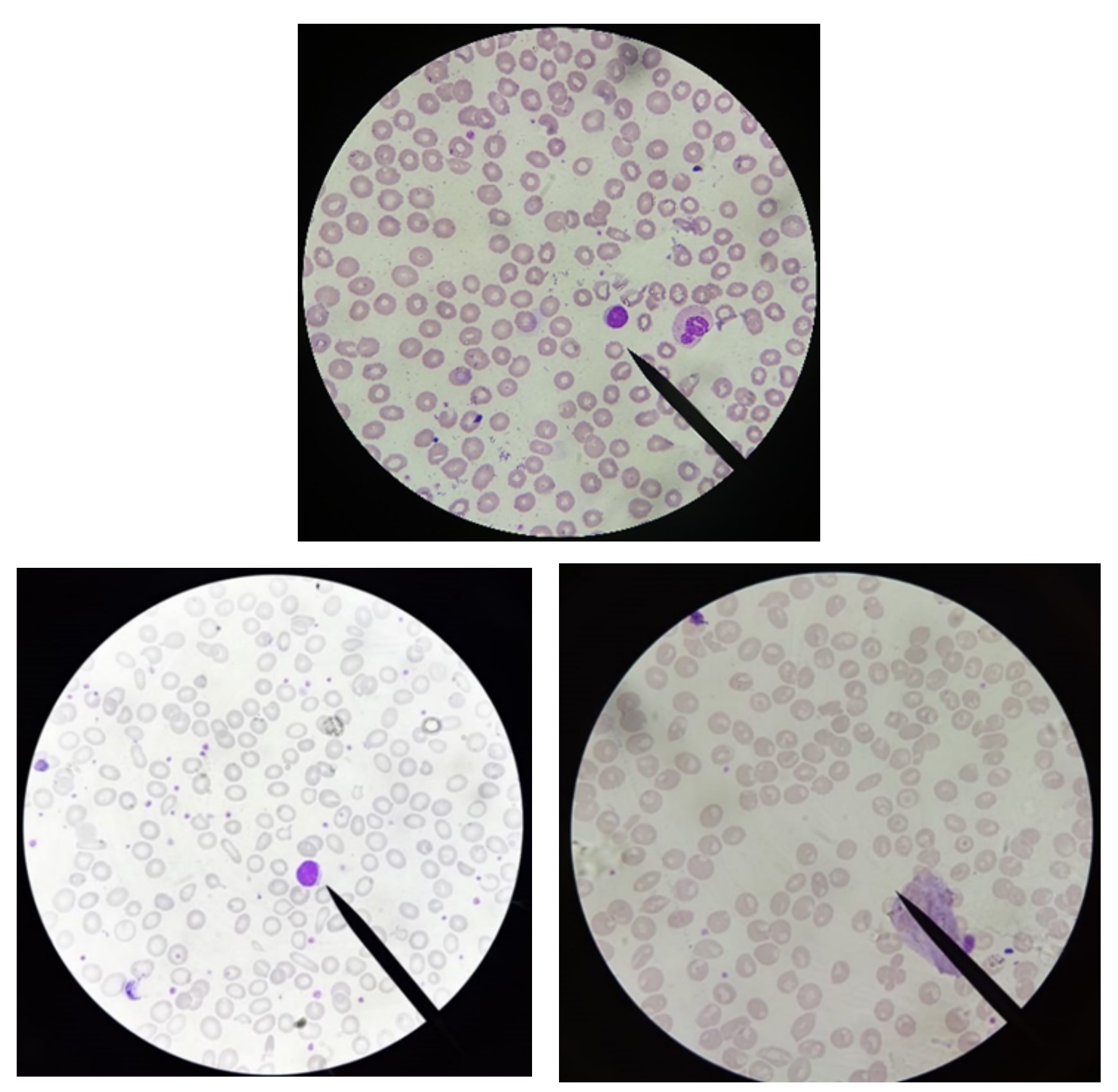
Anemia is a decrease in the number of erythrocytes in the blood circulation or the level of hemoglobin that is less than normal. The three major body mechanisms that cause anemia are excessive destruction of erythrocytes, blood loss, and decreased erythrocyte production. Based on the Basic Health Research (Riskesdas) in 2013, the prevalence of anemic women of reproductive age (WORA) aged 15-44 years in Indonesia was 35.3%. Anemia is classified based on the morphology of erythrocytes, including hypochromic microcytic, normocytic normochromic, and macrocytic. Erythrocyte morphology can be observed using peripheral blood smear examination. The objective of this study was to determine the morphology of erythrocytes in anemic women of reproductive age. This study belongs to descriptive research. The population of the study was 136 women of reproductive age, covering the students of D-IV in Medical Laboratory Technology at Setia Budi University. Forty-one respondents suffering from anemia were taken using a purposive sampling technique. The types of anemia were determined with examination using an Easy Touch hemoglobinometer with the Hb level of less than 12g/dL. Preparation of peripheral blood smear examination using EDTA venous blood and stained with Giemsa. Microscopic examination was performed with 1000x objective magnification. The peripheral blood smear reading showed the erythrocyte morphology that includes normocytic normochromic (38 samples or 93%), microcytic hypochromic (three samples or 7%), and poikilocytosis consisting of teardrop cells, target cells, ellipstocytes, and stomatocytes (five samples or 18%). Further study is required to investigate the correlation of erythrocyte index and peripheral blood smear in anemia.
References
Ardianti, D., Triyani, Y., Afgani, A. dan Herawati & R. (2017). Gambaran Morfologi Apus Darah Tepi dan Karakteristik Pasien Anemia di Laboratorium RS Al-Islam Periode Juni − Desember 2016 Characteristics of Anemia Patients from Peripheral Blood Smear Morphology at Clinical Pathology Laboratory of Al-Islam Hospital ove, Bandung Meeting on Global Medicine & Health (BaMGMH), 1(22), pp. 127–130.
Aulia, G., Udiyono, A., Saraswai, L & Adi, M. (2017). ‘Gambaran Status Anemia pada Remaja Putri di Wilaya Pegunungan dan Pesisir Pantai (Studi di SMP Negeri Kecamatan Getasan dan Semarang Barat)’, Jurnal Kesehatan Masyarakat, 5, pp. 193–200.
Basith, A., Agustina, R. & Dani, N. (2017). Carnitine acyltransferases and their influence on CoA pools in health and disease, Molecular Aspects of Medicine, 5(5–6), pp. 475–493.
doi: 10.1016/j.mam.2004.06.002.
Depkes (Departemen kesehatan RI). (2004). Wanita Usia Subur 2004. Departemen Kesehatan. Jakarta.
Depkes (Departemen kesehatan RI). (2006). Wanita Usia Subur 2006. Departemen Kesehatan. Jakarta.
Depkes (Departemen kesehatan RI). (2008). Anemia pada Wanita Usia Subur 2008. Departemen Kesehatan. Jakarta.
Deshpande, N., Karva, D.,Agarkhedkar, S. & Deshpande, S., (2014). Prevalence of anemia in adolescent girls and its co-relation with demographic factors. International Journal of Medicine and Public Health, 3(4), p. 235. doi: 10.4103/2230-8598.123426.
Gandosoebrata, R. (2016). Penuntun Laboratorium Klinik. Dian Rakyat. Jakarta.
Kiswari, R. (2014). Hematologi dan Transfusi. Erlangga. Jakarta.
Masrizal. (2007). Studi Literatur Anemia Defisiensi Besi. Jurnal Kesehatan Reproduksi, ll(1), pp. 140-145.
Puslitbangkes (Pusat Penelitian dan Pengembangan Upaya Kesehatan Masyarakat, S. dan Litbangkes, B). (2016). Prevalensi dan Faktor Risiko Anemia pada Wanita Usia Subur di Rumah Tangga Miskin di Kabupaten Tasikmalaya dan Ciamis Provinsi Jawab Barat, Jurnal Kesehatan Reproduksi, 7(2), pp. 71–82.
Riskesdas (Riset Kesehatan Dasar). (2007). Badan Penelitian dan Pengembangan Kesehatan. Laporan Nasional Riset Kesehatan Dasar (Riskesdas) Nasional. Jakarta.
Rikesdas (Riset Kesehatan Dasar). (2013). Badan Penelitian dan Pengembangan Kesehatan. Laporan Nasional Riset Kesehatan Dasar (Riskesdas) Nasional. Jakarta.
Riswanto. (2013). Pemeriksaan Laboratorium Hematologi. Alfamedia dan Kanal Media. Yogykarta.
World Health Organization. (2015). The Global Prevalence of Anemia in 2008. Genave.